



:::

Academia Sinica highly values academic freedom and freedom of speech and encourages our colleagues to provide opinions on and propose solutions to key social issues. Nonetheless, research findings and opinions expressed independently by our colleagues do not necessarily reflect the official position of Academia Sinica. We expect all colleagues to adhere to academic norms and take responsibility by citing sources and ensuring accuracy when publishing independently. Research findings and opinions provided on behalf of Academia Sinica should be published according to established procedures.

2024-06-07
  • Research Findings
  • Institute of Cellular and Organismic Biology
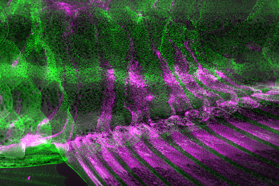
Whole-body Replacement Mechanism Generates Adult Muscle Tissues in the Zebrafish Model

Certain insects, like beetles and butterflies, can undergo massive tissue replacement during their development, a transformation process considered unique to insects. Compared to insects, vertebrates exhibit relatively limited changes in appearance during post-embryonic development. Nonetheless, at the cellular level, whether similar drastic changes occur akin to those observed in insect development remains an unexplored research topic. Dr. Chen-Hui Chen and his team at the Institute of Cellular and Organismic Biology (ICOB) created the palmuscle myofiber tagging and tracking system for in toto monitoring of the growth and fates of ~5000 fast myofibers in developing zebrafish larvae. They found that zebrafish, as a vertebrate model, can similarly undergo complete muscle elimination and replacement during post-embryonic development. Due to limitations in research tools, past studies on vertebrate muscle cells have primarily focused on cell culture models or histological investigations. This study may represent the first instance of simultaneously and continuously observing all muscle cells within an individual at both the organismal and cellular levels. This study finding challenges the current understanding of vertebrate development. This unexpected finding was published in June this year in the EMBO Journal. The first author, Uday Kumar, is affiliated with the Taiwan International Graduate Program, jointly administered by Academia Sinica and National Chung-Hsing University. Other team members include Chun-Yi Fang, Hsiao-Yuh Roan, Shao-Chun Hsu, Chung-Han Wang, and Chen-Hui Chen. This study is supported by funding from the Institute of Cellular and Organismic Biology and grants from Academia Sinica to C.-H.C. (AS-CDA-109-L03 and AS-GCS-112-L01); and grants from National Science and Technology Council, Taiwan, to C.-H.C. (NSTC 110-2628-B-001-016, and NSTC 111-2628-B-001-026).

2024-04-26
  • Research Findings
  • Institute of Molecular Biology
Unraveling how a predatory fungus sense its nematode prey

The capability to sense and respond to the environment is essential for survival and fitness in all organisms. Recent studies have shown organisms from different kingdoms, such as fungi, plants, and mammals can recognize and respond to conserved nematode pheromones, named ascarosides. However, the mechanisms underlying cross kingdom perception of nematode pheromones remain unclear. A research team led by Dr. Yen-Ping Hsueh at the Institute of Molecular Biology, Academia Sinica, revealed that two families of GPCRs are responsible for ascarosides detection, leading to the activation of the cAMP-PKA pathway for trap development in nematode-trapping fungus Arthrobotrys oligospora. The expansion of GPCRs in A. oligospora may have been advantageous for recognizing diverse nematode-derived signals to ensure robust prey recognition during co-evolution. Moreover, the identification of ascarosides receptors in a fungal species sheds light on the molecular mechanisms of ascaroside-mediated cross-kingdom communication. This research has been published on April 22, 2024 in Nature Microbiology. The lead author of this research, Chih-Yen Kuo, is a TIGP-MCB student at Academia Sinica, and collaborators of this research includes Dr. Frank Schroeder at Cornell University and Dr. Yu-Chu Chang at Taipei Medical University. Funding was provided by Academia Sinica and the National Science and Technology Council.

  • :
  • /499