




 Home
Date: 2026-10-22
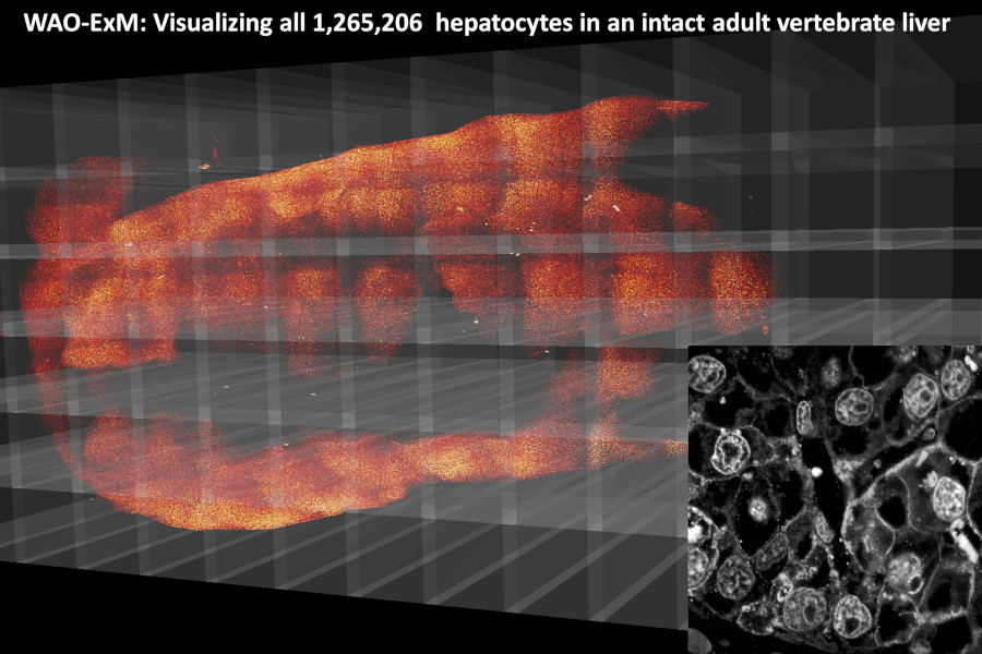
Vertebrate organs undergo massive growth during the post-embryonic period. Yet, our understanding of how this organ-wide process is organized at single-cell resolution has been limited by an inability to monitor individual cells of different types throughout intact adult organs. Here we establish an integrated workflow of whole adult-organ expansion microscopy (WAO-ExM) that enables in toto single-cell-resolved visualization of every hepatocyte within a complete adult vertebrate liver. We found that an intact adult liver spanning ~5 mm thickness had an average of 1,265,206 hepatocytes. Integration of lineage tracing with WAO-ExM revealed that cell number increases were driven by a few hepatocytes undergoing drastic clonal expansion at the whole-organ scale. Disruption of extracellular matrix laminins decoupled liver shaping from total cell number increases, suggesting independent regulatory control of tissue architecture and cell proliferation. Altogether, these findings bridge micrometer-scale cell behaviors with centimeter-scale organ growth, and establish a generalizable platform for adult vertebrate organs to be fully resolved at bona fide single-cell resolution.
-
Link