


 Home
HomeDate: 2026-01-29
Membrane fusion is essential for viral entry. Unlike class I–III fusion proteins, vaccinia virus employs a multi-component Entry Fusion Complex (EFC). Using cryo–electron microscopy, we determined the full-length structure of the vaccinia EFC at near-atomic resolution, revealing a 15-protein asymmetric assembly organized into three layers. The central A16/G9/J5 heterotrimer forms the fusion core, stabilized by conserved PXXCW and Delta motifs, and anchors two A28/H2 adaptor dimers linked to peripheral G3/L5/A21/O3 scaffolds. Structural and evolutionary analyses identify a conserved N-terminal domain in A16 containing a myristoyl-binding pocket and a phenylalanine-rich region that stabilizes the trimer and may regulate lipid engagement. An additional component, F9, binds peripherally to J5, A21, and H2 through Delta-like motifs, reinforcing the prefusion architecture. These findings define the vaccinia EFC as a distinct class of viral fusion machinery and establish a structural framework for understanding poxvirus entry and membrane fusion.
This research was funded by the Academia Sinica Infectious Diseases Research (IDR) Project and National Science and Technology Council (NSTC) Research Project Grants. The co-first authors are IMB postdoc Dr. Chang Sheng-Huei Lin and technician Ching-An Li. Other coauthors include Dr. Chun-Hsiung Wang, facility manager at ASCEM, Dr. Meng-Chiao Ho, Associate Research Fellow, and postdoc Dr. Min-Chi Yeh at IBC, Dr. Hsin-Ming Lee, Associate Research Fellow, and postdoc Dr. Hua-De Gao at IoC, Hsiao-Jung Chiu, a TIGP-MCB Ph.D. student, and Dr. Chi-Fei Kao, Assistant Professor at the School of Veterinary Medicine of NTU. The corresponding authors are Dr. Wen Chang, Research Fellow, and postdoc Dr. Chang Sheng-Huei Lin at IMB. The research findings were published in Science Advances on January 14, 2026, and were selected as a Research Highlight of the journal.
-
Link
-
 Cryo-EM Structure of the Vaccinia Virus Entry Fusion Complex Reveals a Multicomponent Fusion Machinery

-
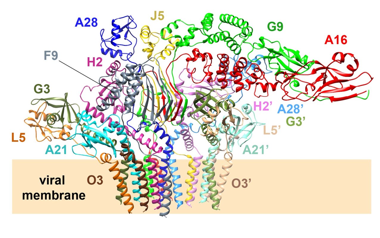
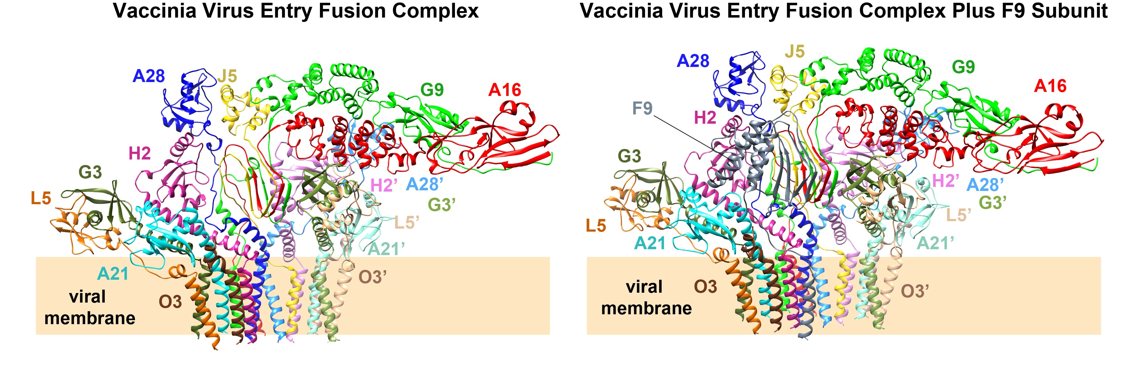 The figure presents the cryo-EM structures of the poxvirus entry fusion complex (EFC,left panel) and EFC plus F9 subunit (right panel), revealing amultiprotein fusion machinery. The EFC is a 15-protein assembly composed of one copyof A16, G9, and J5 forming the central trimer, two copies of A28 and H2 flanking bothsides of the central trimer, and two peripheral sets of G3, L5, A21, and O3 at the outermost region of the complex. With overall dimensions of approximately 210Å ×137Å × 108Å, the EFC adopts a flower-bouquet-like architecture, in which thetransmembrane helices resemble a bundled stem, and the ectodomains extend outward like a blooming cap.Photo credit: Academia Sinica.
